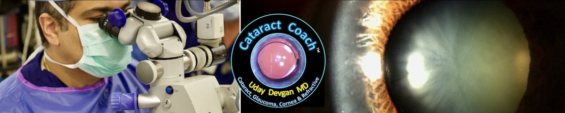


3094: cataract surgery with only a half view

Operating through dense corneal scarring and lipid keratopathy requires adapting surgical maneuvers to rely heavily on the clear half of the cornea for intraocular visualization. Perhaps staining the anterior capsule with trypan blue could enhance tissue contrast however our guest surgeon is able to create a beautiful capsulorhexis without it. To maintain safety when nuclear disassembly extends under the opaque corneal zone, the surgeon must utilize muscle memory and years of experience to prevent inadvertent capsule or iris trauma. Elevating nuclear fragments into the clear optical zone before emulsification gives precise, controlled phacoemulsification. Watch the video to see how careful instrument positioning can overcome partial corneal opacity during surgery. How do you adapt your visualization and surgical approach when operating through localized corneal scars or lipid deposits? Please comment your thoughts below.
