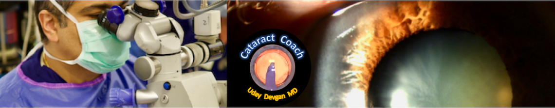


The phaco probe is placed in the eye at the beginning of the cataract surgery and when ultrasonic energy was applied, a white plume of material appeared at the tip. What is this material? Is there a danger here? Will it cause an infection or inflammation? What is your next step?
Click below to learn from this cataract quiz about this white plume:
It looks like the phaco tip was completely clogged, or the aspiration tube was folded, or the aspiration malfunctioned. When there’s no aspiration the emusified lenticular matter comes in the anterior chamber, mostly in very hard nuclei. Normally it disperses, but in this eye the white cloud did not because the anterior chamber was filled with OVD. Maybe even both the aspiration and the irrigation were blocked, as there is no egress of BSS visible neither through the main port nor via the side port when the phaco is activated the first time. So the white material did not come out of the phaco tip but arose in front of it.
Thank you for the keen insight
I have had this exact thing happen multiple times and have figured out it is related to activating phaco power directly into dispersive OVD on a low flow, low vacuum setting (sculpt). Notice the AC was completely refilled with likely dispersive OVD here and phaco was activated right in the material. When i switch the phaco to chop or quad removal with high flow, high vacuum then this does not happen. Similarly, if i do not refill the AC with dispersive OVD after hydrodissection and prior to phaco, then this does not happen. This never happened to me until I started taking the extra step of refilling the AC again with dispersive OVD immediately prior to phaco. My remedy is to either use a little less dispersive during this step, or to start with high flow, high vacuum phaco mode and remove some of the anterior epinuclear material along with some of the dispersive OVD that is directly overlying the lens before sculpting a groove.
Excellent observation. I also do the same – just a small aliquot of dispersive OVD against central corneal endothelium prior to inserting the phaco probe.